
Happy Tuesday, friends! Building on yesterday's Medical Monday about the anatomy of the Aorta, today's Topic Tuesday will dive into the Aortic Arch.
What is the Aortic Arch?
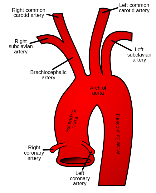
The Aortic Arch is the section of the aorta that lies between the Ascending and Descending aorta, making a 180 degree turn. It features a Greater Curvature (the outer curvature) and a Lesser Curvature (the inner curvature. Its most important feature is the 3 blood vessels the branch off from the Greater Curvature- the Brachiocephalic Artery, Left Common Carotid Artery, and Left Subclavian Artery.
Brachiocephalic Artery: This is the first vessel to branch off of the arch. It quickly divides into 2 arteries, the Right Subclavian Artery and the Right Common Carotid Artery. As you may have noticed from their names, these 2 arteries are the right-sided counterparts to the other Aortic Arch branches. The Right Subclavian artery mainly supplies the blood to the right arm, while the Right Carotid artery mainly supplies blood to the head.
Left Common Carotid Artery: This artery is the second of the 3 branches off of the arch and also supplies the head, similarly to the Right Common Carotid.
Left Subclavian Artery: The last branch off of the arch, this artery supplies the blood to the left arm.
Clinically, these 3 branches provide blood flow to a significant portion of the body, most importantly the brain. Most of the brain (80%) is supplied by branches of both the Left and Right Common Carotid Artery (known as the Internal Carotid Arteries), so clots or narrowing of these arteries can cause strokes. Smaller branches of both the Left and Right Subclavian artery (known as the Vertebral Arteries) supply a smaller portion (20%) of the brain and can also be responsible for strokes.
All of these vessels that supply the brain connect to each other in something called the Circle of Willis, which can be helpful when blood flow through one of the vessels is compromised since it may find a detour and continue to supply the entire brain. Unfortunately, this isn't always a perfect solution as some people have variable Circle of Willis anatomy.

What else can the Aortic Arch do?
Beyond just being a fancy bend in the aorta that carries blood, the Aortic Arch also contains baroreceptors. Baroreceptors sense the blood pressure based on the stretch in the aortic walls and send signals to the brain when things are out of balance. If the pressure is too high, the brain can signal for the walls of the arteries to relax (vasodilation) and drop the blood pressure. The brain can also signal for the heart rate to drop and decrease the contraction of the heart.
Navigating the Aortic Arch during surgery
The repair of aortic aneurysms and dissections often requires operation on the arch or beyond, which poses a significant challenge. While the heart-lung machine can usually be used to bypass the part of the heart and aorta being operated on, it is much more difficult to bypass the important branches of the arch. In order to keep blood flowing to the brain, Selective Cerebral Perfusion is sometimes used. This allows for blood flow to the rest of the body to be stopped while the arch is operated on and the brain receives a separate flow of blood.
Here is how it works:
The body is cooled down to slow its metabolism and protect the tissues.
Surgeons place tubes into one of the aortic arch branches and pump blood forwards to selectively give the brain oxygen while the rest of the body receives no flow.
The aortic arch can be repaired and the selective cerebral perfusion stopped once the repair is completed, and then the heart-lung machine can resume flow to the entire body.
Phew, that was a lot, but I hope you can appreciate the complexity of the Aortic Arch and why it deserves its own post! Stay on the lookout for future content and thanks again for tuning in to this week's Topic Tuesday!
-Purab
More Reading:
Comments