It's Topic Tuesday!
- Alvaro Jose Martinez Santacruz
- Nov 12, 2024
- 3 min read
Updated: Nov 19, 2024
It's Topic Tuesday!
Welcome, everyone! I hope life has been treating you well. Today, we’re diving into coarctation of the aorta (CoA), a condition that accounts for about 5% of all congenital heart defects and is often diagnosed in children and adults under 40.
What is the coarctation of the aorta (CoA)?
It is a congenital heart defect characterized by a narrowing of the aorta and can be compared to a pipe. Just as a narrow pipe restricts the flow of water, making it difficult to reach its destination, a narrowed aorta restricts blood flow, creating challenges for the heart to pump effectively and supply the body with oxygenated blood.

Credits: Coarctation of the aorta. Medlineplus.gov n.d.
What are the symptoms?
It will depend on the degree of narrowing because this will directly affect blood flow, so the narrower, the more symptoms.
Neonates (Early Stage): Patients may often be asymptomatic, but can exhibit symptoms such as rapid breathing, poor feeding, weak leg pulses, and lower oxygen levels in the legs compared to the arms. They may also experience heart failure.. An echocardiogram typically shows a narrowed aorta
Neonates (Late Stage): Symptoms can include rapid breathing, poor feeding, weak leg pulses, and a heart murmur. An echocardiogram may reveal thickened heart walls and signs of heart strain, while a chest X-ray may show an enlarged heart and fluid accumulation in the lungs.
Children to Adults: These patients may experience headaches, nosebleeds, and leg pain during exercise. Other signs can include high blood pressure (notably a difference between arm and leg measurements) and a continuous heart murmur. An echocardiogram may indicate heart strain, along with the potential for rare complications such as aortic tears.
Evaluation
During a physical exam, the pulse in the groin (femoral) area or the feet may be weaker than the pulse in the arms or neck (carotid). Sometimes, you may not be able to feel the femoral pulse at all.
The most common exam to diagnose this condition is echocardiography, which can also help monitor the patient after surgery. Older children may need a heart CT, and an MRI or MR angiography of the chest may also be necessary.
Treatment
Surgery
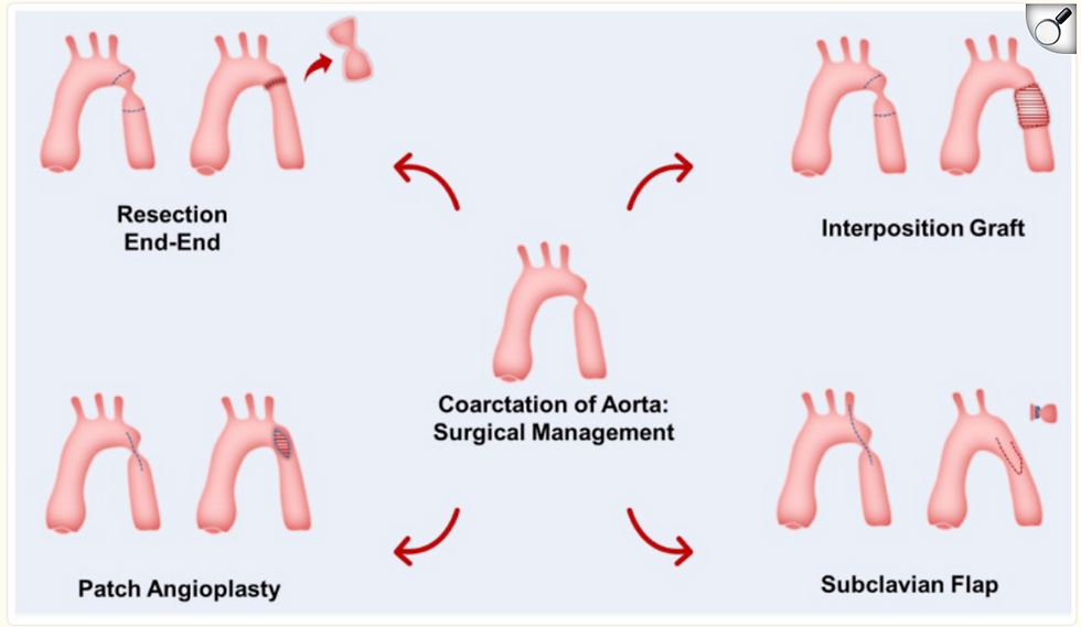
There are several surgical options for repairing CoA. One common approach is end-to-end anastomosis, where the segment of the aorta that is narrowed (coarctation) is removed, and the ends of the aorta are sutured together. Another technique is subclavian flap repair, which involves cutting out the subclavian artery (one of the vessels that classically come off the aortic arch) andusing the flap from the subclavian artery to replace the narrowed section the aorta.

Credits: Raza S, Aggarwal S, Jenkins P, Kharabish A, Anwer S, Cullington D, et al. Coarctation of the aorta: Diagnosis and management. Diagnostics (Basel) 2023;13:2189.
Fun Fact: The anatomist Morgagni described CoA of the aorta in 1760
That concludes our session for today, everyone. I am Alvaro, a medical student, and I hope you found this informative. I'm delighted to have joined you for this Topic Tuesday.
Bibliography
[1] Law MA, Collier SA, Sharma S, Tivakaran VS. Coarctation of the aorta. StatPearls, Treasure Island (FL): StatPearls Publishing; 2024.
[2] Raza S, Aggarwal S, Jenkins P, Kharabish A, Anwer S, Cullington D, et al. Coarctation of the aorta: Diagnosis and management. Diagnostics (Basel) 2023;13:2189. https://doi.org/10.3390/diagnostics13132189.
[3] Coarctation of the aorta. Medlineplus.gov n.d. https://medlineplus.gov/ency/article/000191.htm (accessed November 9, 2024).

Opmerkingen